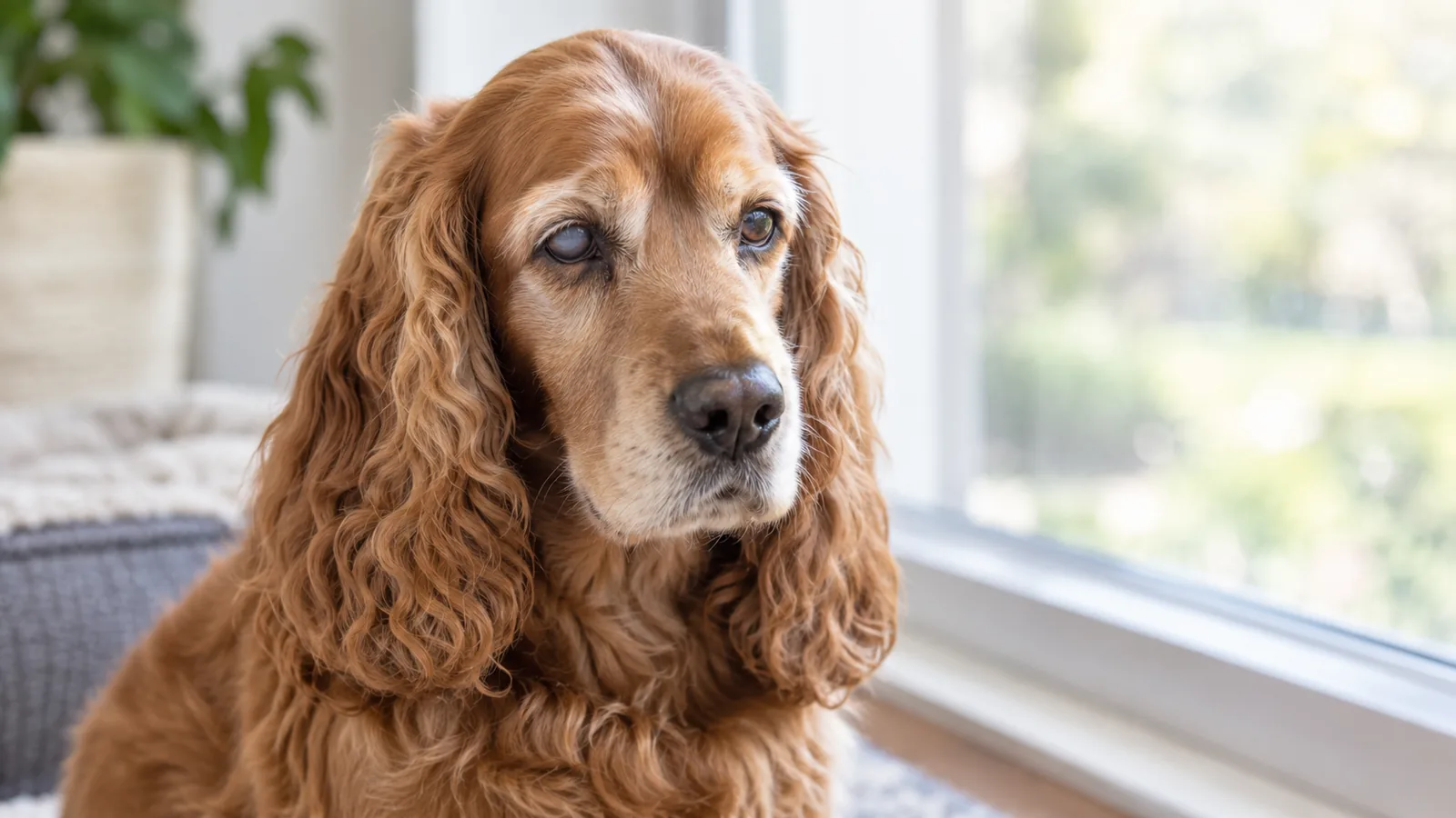
Your dog wakes up squinting. One eye is red, slightly cloudy, and clearly bothering him. By afternoon the eye looks bigger than the other one, he's pawing at his face, and he won't let you near it. This is what acute glaucoma looks like, and it's one of the few eye conditions where the time between "something's off" and "permanent blindness" can be measured in hours.
Glaucoma develops when fluid that's supposed to drain out of the eye can't leave at a normal rate. Pressure builds inside the eye, and that pressure damages the optic nerve and retina. The damage is largely irreversible, which is why acute glaucoma is a true emergency rather than a watch-and-wait. Chronic glaucoma is the slower-moving cousin, often caught on a routine wellness exam before vision loss is obvious. Either form needs the same fundamental thing: accurate diagnosis through eye pressure measurement, followed by a plan focused on dropping the pressure, controlling pain, and saving whatever vision is still there to save.
At Noah's Ark Veterinary Hospital in Williamsburg, we measure intraocular pressure in-house and evaluate eye emergencies promptly during our hours, with coordinated referral to a veterinary ophthalmologist for cases that need advanced surgical management. Our emergency services handle acute eye presentations, and our wellness exams include the eye assessment that catches chronic disease before it becomes a crisis. If your dog or cat has a red, cloudy, painful, or enlarged eye, don't wait for a routine slot. Contact us right away, or head to the nearest 24-hour emergency hospital if we're closed.
Glaucoma in Pets: Key Points
- Acute glaucoma is an emergency. Permanent vision loss can occur within hours.
- The eye looks and feels wrong: typically red, cloudy, enlarged, and painful. Your pet may squint, paw at the face, or hide.
- Pressure has to be measured. Diagnosis requires eye pressure measurement (tonometry), not just looking at the eye.
- Treatment has three goals: dropping pressure, controlling pain, and preserving whatever vision remains.
What is glaucoma and what causes it?
Inside a healthy eye, fluid called aqueous humor is constantly produced and drained at matching rates, keeping the internal pressure steady. When that drainage gets blocked or slowed down, fluid backs up and pressure rises. High pressure damages the optic nerve (the cable carrying visual signals from the retina to the brain), and once that damage is done, it doesn't undo.
Glaucoma falls into two broad categories:
| Type | Underlying cause | Typical course |
| Primary canine glaucoma | An inherited abnormality of the eye's drainage angle, seen in predisposed breeds (Cocker Spaniels, Basset Hounds, Beagles, Bostons, Chow Chows, Akitas, and others) | Usually starts in one eye and progresses to the other within 8 to 12 months |
| Secondary glaucoma | Another eye problem: inflammation, trauma, cancer, or chronic eye disease | Sometimes resolves if the underlying problem is treated early; becomes permanent if treatment is delayed |
Glaucoma in cats
Glaucoma in cats is a different animal, both literally and clinically. Cats almost never get primary glaucoma. Instead, feline glaucoma is almost always secondary to something else going on inside the eye, most often chronic uveitis (inflammation), and it tends to develop more slowly than the dramatic acute presentations we see in dogs.
The signs can be subtle: a slightly larger or off-color eye, a pupil that doesn't quite match the other side, a cat who's a little less interactive than usual. Because the presentation is quieter, cat glaucoma is often more advanced by the time it's diagnosed, which is why any noticeable change in a cat's eye is worth a visit even if she's not in obvious distress.
Triggers of secondary glaucoma
Secondary glaucoma usually develops because something else going on inside the eye, often uveitis (inflammation inside the eye), disrupts the normal drainage of fluid. The list of things that can trigger uveitis is long, and many of the causes are systemic diseases that affect the eye as one of several organs involved. Identifying and treating the underlying cause is part of treating the glaucoma.
In dogs, common triggers of uveitis include:
- Tick-borne diseases like Lyme disease, ehrlichiosis and anaplasmosis, all of which can cause eye inflammation as part of systemic disease
- Bacterial diseases like leptospirosis and canine brucellosis
- Viral diseases like canine distemper
- Heartworm disease, which can occasionally produce ocular signs
In cats, common triggers of uveitis include:
- Viral diseases like feline immunodeficiency virus (FIV), feline infectious peritonitis (FIP), and feline leukemia (FeLV)
- Bacterial diseases like bartonella, caught from fleas
Triggers that affect both dogs and cats include:
- Fungal diseases like blastomycosis, histoplasmosis, and cryptococcus
- Parasitic infections like toxoplasmosis
- Eye injuries, especially corneal ulcers and displaced lenses
- Systemic hypertension, often secondary to kidney disease or thyroid disease
- Diabetes
- Eye tumors like lymphoma and melanoma
- Cataracts
This is part of why a workup for secondary glaucoma often includes more than just an eye exam. Bloodwork, infectious disease testing, blood pressure measurement, and sometimes imaging help us figure out what's driving the inflammation. Treating the eye without addressing the underlying cause rarely produces lasting control. Our hospital has the capability to run the blood tests and other testing needed to find the root cause.
What are the signs of glaucoma in pets?
There are really two different presentations to know about: the acute crisis and the slower chronic version. Both need attention, but the urgency is different.
Acute glaucoma: the emergency presentation
Acute glaucoma hits fast and hits hard, and is *painful*. Most or all of the following will be present:
- A noticeably red eye
- A cloudy or hazy cornea (often with a bluish tint)
- An enlarged eye (more obvious in dogs)
- A dilated pupil that doesn't respond to light
- Squinting or holding the eye closed
- Excessive tearing
- Pawing at the face or rubbing the eye on furniture
- Bumping into things or hesitating in familiar areas
- Hiding, lethargy, or other behavior that doesn't fit
- Vocalization in some cases
Acute glaucoma symptoms progress from "something's off" to "permanent vision loss" in hours, not days. The window for saving vision is small, and it doesn't reopen.
What to do right now if you see acute glaucoma
- Don't wait for a routine appointment slot. Call us as you're heading out the door for an emergency visit.
- If we're closed, head to the nearest 24-hour emergency hospital. This is what they're there for.
- Don't put anything in the eye unless we tell you to. Some over-the-counter eye products can make things worse.
- Keep your pet calm and comfortable in the car. Pain plus stress isn't helpful, but the trip is worth it.
Chronic glaucoma: the slower version
Chronic glaucoma develops more gradually, and it often presents differently. The eye may be enlarged from longstanding pressure (a condition called buphthalmos), but the acute pain has subsided as the eye has adapted. Vision is typically already gone by the time owners notice the change. Subtle signs to watch for:
- An eye that's gradually bigger than the other
- Mild cloudiness that doesn't seem to be getting worse
- Reduced interest in toys or activities that require sharp vision
- Hesitation on stairs or in low light
- A slightly off-color cornea
The cat or dog who's been mildly bumping into things at night for a few months and now has an eye that "doesn't quite look right" may have chronic glaucoma that's been building for a while. The conversation here isn't usually about preserving vision; it's about controlling pain and figuring out the right long-term plan.
How is glaucoma diagnosed?
The defining test is tonometry, which is a quick, painless measurement of pressure inside the eye. We do it in-house at the time of the exam. Normal pressure in dogs and cats sits roughly between 10 and 25 mmHg. Glaucoma diagnoses typically involve pressures over 30, and acute crisis cases can run 50, 60, or even 70+ mmHg. Here's the thing: the eye can look only mildly abnormal while the pressure is dangerously high. The number on the tonometer matters more than what the eye looks like from the outside.
A complete glaucoma workup typically includes:
- Tonometry to measure eye pressure
- Fundic examination to look at the optic nerve and retina for damage
- Slit lamp biomicroscopy for a detailed look at the front of the eye
- Gonioscopy to evaluate the drainage angle (usually done by an ophthalmologist when surgery is being considered)
- Ocular ultrasound when the cornea is too cloudy to see through (also typically done at an ophthalmologist)
A thorough eye exam also rules out the conditions that mimic glaucoma, especially corneal ulcers, uveitis, cataracts, and lens luxation. Catching chronic and at-risk presentations at wellness visits, before pressure has done its damage, is one of the strongest cases for routine eye exams in older pets and in predisposed breeds.
How is glaucoma treated with medication?
Treatment goes after three things at once: bringing the pressure down, controlling pain, and preserving whatever vision is left. Medical management for primary glaucoma is lifelong, and the unaffected eye also needs preventive treatment because primary glaucoma typically catches up to the other eye within 8 to 12 months. Pressure checks on a regular schedule confirm the medications are doing their job. The drops themselves are typically small-volume, given once or twice daily, and most pets accept them with practice. We'll walk you through how to administer eye medications at the visit, because correct technique is the difference between drops that actually reach the eye and drops that run down the side of your pet's face.
A few practical points families often ask about:
- Some glaucoma drops sting briefly. Your pet may shake her head or squint after a drop. This is normal, brief, and not a reason to skip doses.
- The bottles often look identical. Label them clearly with the eye and the schedule, especially when more than one medication is in rotation.
- Don't double-dose if you miss one. Give the next dose at the regular time and move on.
- Storage matters. Some eye medications need refrigeration; check the label.
When is surgery needed for glaucoma?
Surgery enters the conversation when medications can't keep pressure controlled, when an eye is blind and painful and comfort has become the priority, or when specific anatomy points toward a surgical fix. Surgical management of glaucoma options include:
- Cyclophotocoagulation uses a laser to reduce fluid production in the eye
- Gonio-implant placement creates a drainage shunt to redirect fluid out of the eye
- Intrascleral prosthesis is a cosmetic option for some blind painful eyes
Most of these procedures require referral to a board-certified veterinary ophthalmologist, and we coordinate that referral when surgery is on the table. For end-stage cases, enucleation often produces the dramatic comfort improvement your pet has been needing. The eye has already lost vision; the goal shifts from saving sight to ending chronic pain. Pets adapt remarkably well to having one eye or none, and their quality of life after enucleation is consistently better than living with an eye that hurts every day. We perform enucleations for dogs and cats, and will answer all of your questions about keeping a blind pet living comfortably.
What does long-term glaucoma care look like?
Ongoing care for a pet with glaucoma usually includes:
- Regular eye pressure checks, every 2 to 4 weeks at first and then every 1 to 3 months once things are stable
- Watching for changes in eye appearance, comfort, or behavior at home
- Medication adjustments as the disease evolves
- Preventive treatment of the unaffected eye in primary glaucoma cases
- Pain assessment at every visit
Glaucoma is often progressive even with optimal management, and the conversations that happen along the way matter as much as the medications. Most families end up walking through several "what now" moments: when to escalate medications, whether to refer to ophthalmology, when surgery becomes the right move, when to focus on comfort rather than vision. We'll have these conversations honestly, including when the most loving option is removing a painful eye.
How can I support a pet with vision loss?
Pets adapt to vision loss extraordinarily well, especially when the loss has been gradual. The world becomes a map of sounds, smells, and remembered routes, and most blind or low-vision pets are confidently navigating their homes within weeks of losing sight. Muffin’s Halo is a great resource for living with a blind pet, and they offer halo attachments for harnesses that keep your pet from bumping into objects.
Most families end up surprised by how well their pet adapts. The dog who couldn't possibly figure out the back yard finds her favorite sniffing spots within days. The cat who seemed lost in the living room is back to claiming his sunbeam by week two. The adjustment is real, but it's not a tragedy. It's a transition, and pets are good at it.
Common questions about glaucoma in pets
How can I tell if my pet's eye problem is glaucoma versus something else?
Honestly, you usually can't tell from the outside. Many eye problems cause similar redness and cloudiness, and the only way to confirm glaucoma is to measure the pressure. The combination of a red, cloudy, painful eye warrants prompt evaluation regardless of which specific condition is suspected. Don't try to diagnose at home. Come in and let us check the pressure.
My breed is at risk for glaucoma. Should I screen proactively?
For breeds with strong primary glaucoma predispositions, periodic eye pressure screening starting in middle age is reasonable. We'll talk through the specific recommendation based on your pet's breed and family history. For at-risk breeds, the simple act of measuring pressure at wellness visits often catches changes early enough to matter.
Will my dog or cat lose vision even with treatment?
Often, eventually. Glaucoma is typically progressive, and the goal of treatment shifts over time from preserving vision to maintaining comfort. Many pets retain useful vision in at least one eye for years with appropriate management, and early detection plus aggressive treatment give the best chance of preserved vision. Even when vision is ultimately lost, well-managed glaucoma means a comfortable pet, not a suffering one.
What does enucleation actually involve?
Surgical removal of the affected eye, performed under general anesthesia. The eyelids are typically closed and sutured permanently, producing a smooth closed eyelid where the eye used to be. Recovery is usually straightforward, with most pets back to normal activity within 1 to 2 weeks. The end result is a pet who's no longer experiencing chronic eye pain and who adapts well to vision from one eye. For end-stage glaucoma, this often produces the comfort improvement the pet has been needing for months.
Can glaucoma be prevented?
Primary glaucoma is inherited, so it can't really be prevented. What you can do is screen at-risk breeds proactively, take any change in your pet's eyes seriously, and treat conditions like uveitis or eye injuries promptly to reduce the risk of secondary glaucoma developing.
My pet has one eye that's already blind from glaucoma. How worried should I be about the other eye?
In primary glaucoma, very worried, and we'll be treating the second eye preventively for exactly that reason. The other eye typically follows within 8 to 12 months without preventive treatment. With preventive drops, we can often delay or sometimes prevent the second eye from being affected.
Protecting your pet's eye health
Glaucoma is one of those conditions where the timeline matters more than almost anything else. Pets whose families recognize the early signs and act fast keep more vision and comfort than pets whose presentation gets watched at home for a day before coming in. For pets already diagnosed, faithful daily medications and consistent rechecks are what make the difference between years of comfortable vision and a hard, fast progression.
If your pet shows any sign of eye pain, redness, cloudiness, or sudden vision changes, contact us right away. For acute presentations, our emergency services provide same-day evaluation while we're open, and we'll coordinate emergency referral after hours when needed.