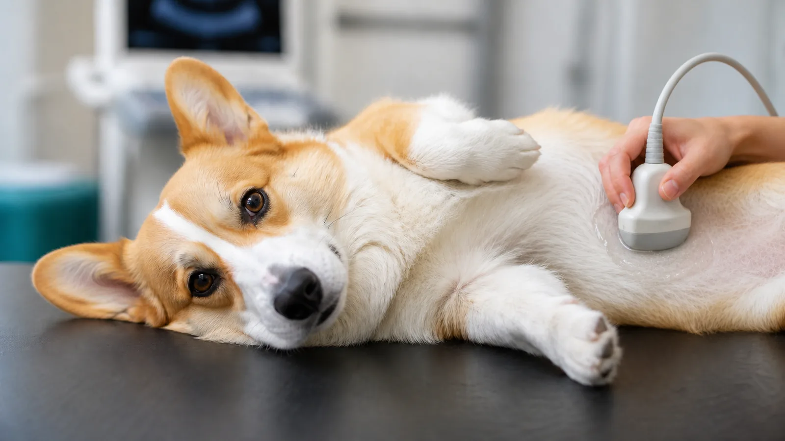
Plenty of families want to know why one pet is sent for an X-ray while another needs an ultrasound, and the reason is that each tool answers a different question. X-rays are best at bones, the chest and lungs, swallowed objects, and the overall size and shape of organs. Ultrasound sees inside soft tissue in real time, which is how we study the layers of an intestinal wall or watch a heart valve move, and it uses no radiation. Neither tool is better, and pets often need both, because each shows what the other cannot. Advanced tools like CT, MRI, and endoscopy go further still on specific problems, and they are the reason some pets are referred for a more detailed view.
Noah's Ark Veterinary Hospital, AAHA accredited and here in Williamsburg, keeps digital X-ray and both abdominal and cardiac ultrasound on site, so the first look at what is going on happens right here with your own veterinarian. When your pet is vomiting, coughing, or simply not acting like themselves, we pair the physical exam with our in-house imaging to narrow down the cause, and if a case calls for CT, MRI, or endoscopy, we help you arrange it. If you are unsure whether your pet's symptoms need imaging, ask us your questions and we will tell you honestly what we would want to see.
Which Scan Answers Which Question
- X-rays and ultrasound aren't ranked against each other, and the one your pet needs depends on which question has to be answered first.
- Many pets need both, because a radiograph shows the outline and position of an organ while ultrasound shows what's happening inside it.
- CT, MRI, fluoroscopy, and PET-CT answer narrower questions, and those scans mean a referral to a specialty or university hospital.
- Endoscopy uses a small camera on a flexible tube to look inside directly, and it can collect a biopsy or retrieve a swallowed object during the same procedure.
What Do X-Rays and Ultrasound Each Do Best?
Every diagnostic imaging decision starts with what has to be seen: bone, gas, and the outline of an organ show up on X-ray, while the layered texture inside that same organ shows up on ultrasound.
| The question | What an X-ray gives you | What an ultrasound gives you |
| Bones, joints, fractures | The primary tool, start to finish | A limited role, mostly soft tissue around a joint |
| Swallowed objects, blockages | Many objects, plus the gas pattern of an obstruction | Detail of the intestinal wall and how it's moving |
| Lungs and airways | The best first look at lung tissue | Helpful when fluid or a mass is present |
| Heart | Size, shape, and position | Valves and muscle moving in real time |
| Liver, kidneys, bladder wall | Size and outline only | Layer-by-layer internal detail |
| Radiation | A very low dose | None, it uses sound waves |
The two work as a pair more often than families expect. A dog with a swollen belly might need a radiograph to rule out a twisted stomach and an ultrasound to check the spleen and liver, and one without the other leaves half the picture blank. It's also worth knowing that a single image doesn't always close the question, since some obstructions, early tumors, and soft tissue changes only appear on ultrasound or on a repeat image the next day. Our in-house digital X-ray and ultrasound cover most everyday questions, so when a case needs both halves, you're not driving somewhere else for the second one.
When Does a Vet Reach for Imaging?
Imaging comes out when the exam raises a question that hands can't settle: a belly that hurts everywhere, a cough that won't quit, a leg that won't take weight. The pattern of signs points us toward a body system, and the body system points toward the tool.
Vomiting and Diarrhea
Repeated vomiting in dogs and cats can trace back to something as ordinary as a scavenged meal or as serious as an intestinal blockage, pancreatitis, or kidney disease, and a physical exam alone can't tell those apart. Radiographs find swallowed objects and the gas pattern that suggests an obstruction, while ultrasound reads the layers of the bowel wall and checks the pancreas, liver, and nearby lymph nodes. When a blockage is suspected but not obvious, a contrast study, where a pet swallows a special dye that outlines the GI tract on X-ray, can show exactly where things stop moving. Repeat vomiting, blood in the stool, or a missing sock on the same day your dog stopped eating all deserve urgent care during our open hours instead of a wait-and-see night.
Coughing and Trouble Breathing
Breathing problems are the ones that shouldn't wait until morning. Open-mouth breathing in a cat, labored effort, or gums that look pale or bluish mean your pet needs to be seen immediately, and it helps to call us before you head over so we're ready at the door. A single set of radiographs covers a lot of ground: lung tissue, the windpipe, and the size and shape of the heart. A cat who crouches low and coughs in a way that looks like a hairball attempt that never produces anything is often showing feline asthma, and chest X-rays are what reveal the thickened, inflamed airways behind it. In small breeds, a dry honking noise can point toward a collapsing windpipe, one of the few problems better watched in motion than in a still image. Coughing at night, tiring quickly on a familiar walk, and a faster breathing rate during sleep are early signs of heart disease, and a chest X-ray measuring heart size is usually the first objective step, with an echocardiogram (cardiac ultrasound) filling in how well the valves and muscle are working.
Limping and Bone or Joint Pain
Lameness is hard to pin down by feel, especially in a dog who braces the moment you touch the sore spot. A dog can still put weight on a fractured leg and a cat can still jump on one, which is why broken bones are confirmed with an X-ray instead of a hands-on exam alone. Radiographs also settle arthritis, hip dysplasia, and bone tumors, and they tell us whether trouble sits inside the joint or in the bone itself. Ultrasound plays a smaller part here, useful for a torn muscle or a swollen tendon in the tissue surrounding a joint. Sometimes a CT or MRI is needed to get a better view of complex fractures or tendons and ligaments. After a fall, a car, or any sudden refusal to bear weight, book a visit rather than reaching for the medicine cabinet.
Dental Disease Below the Gumline
That smell when your dog yawns at you usually starts somewhere you can't see. Most of a tooth sits below the gumline where the eye can't follow, so dental radiographs taken during a cleaning are what turn up root abscesses, fractured roots, and the painful resorptive lesions so common in cats. Ultrasound has no role in the mouth at all, since sound waves can't pass through enamel and bone. Our dental cleanings with digital dental X-rays include a look at every tooth, because a crown that appears perfectly healthy can hide an abscess underneath. Bad breath, drooling, pawing at the face, or chewing on one side only are worth an appointment. In cases where disease is severe or complex, a dental specialist may perform a CT.
Urinary and Reproductive Problems
Straining, frequent small squats, and blood in the urine often point to urinary stones, and while many types show up clearly on an X-ray, others are visible only on ultrasound. Sound waves also read the bladder wall for thickening, check the kidneys for changes in size and shape, and look at the prostate or uterus when something seems off. In an unspayed female who's drinking more and picking at her food a few weeks after a heat cycle, imaging is how we look for pyometra, an infected uterus that turns into an emergency quickly. X-rays confirm a late-term pregnancy once tiny skeletons have formed, and ultrasound picks up a pregnancy far earlier and shows beating hearts. A male cat straining in the litter box without producing urine needs same-day evaluation, and never press on his belly to try to move things along.
Finding and Following Cancer
Finding a new lump is a frightening moment, and plenty of them turn out to be something simple. Much of diagnosing cancer happens through imaging, with chest X-rays checking whether disease has reached the lungs and ultrasound examining the abdominal organs and guiding a needle to exactly the right spot for a sample. Many of those samples are read in house through our cytology, so the conversation can start without a long wait. Once a diagnosis is in hand, repeat scans become part of the ongoing plan, showing whether a mass is shrinking, holding steady, or asking for a different approach. If you've noticed a growing lump, weight coming off without a diet change, or a pet who's tired in a way that doesn't fit their age, our veterinarians will start with an exam and explain what a closer look would tell us.
Why Would Your Vet Refer Your Pet for Advanced Imaging?
A referral usually means the everyday tools answered part of the question and one piece is still missing, not that something has gone wrong. CT, MRI, fluoroscopy, and PET-CT live at specialty and university hospitals, so we arrange the referral and send your pet's images and history along. The specialist runs the scan, and our veterinary team stays in the loop and helps you decide what to do with the answer.
CT Scans
A flat X-ray stacks everything in the body onto a single plane, and a CT scan solves that by combining hundreds of images into a three-dimensional view that separates overlapping structures, which is why it's the tool of choice for complex fractures, nasal disease, and surgical planning. It's also fast, which matters for a patient under sedation.
MRI
For the brain and spinal cord, MRI is the clearest window there is, using magnetic fields rather than radiation to separate the fine soft-tissue detail that answers questions about seizures, sudden weakness, or a disc pressing on the spinal cord. The tradeoff is time, and because the patient has to stay perfectly still, general anesthesia is part of the plan.
Fluoroscopy
Some problems only appear while the body is moving, and fluoroscopy captures them by running X-ray images continuously, like video, so a swallowing disorder or a windpipe that collapses with each breath can be watched as it happens.
PET-CT and Functional Imaging
Where other scans show what tissue looks like, PET-CT shows what it's doing, flagging areas of unusually high metabolic activity that can mark active tumor tissue, which makes it most useful for staging cancer and tracking how a pet is responding to treatment. It's newer in veterinary medicine and offered at only a small number of specialty centers.
What Can Endoscopy See That a Scan Can't?
Endoscopy skips the picture and looks directly, threading a small camera on a flexible tube into the body without surgery. It shows color, texture, and the surface of a lining in a way no scan can reproduce, and during the same procedure it can collect a biopsy or pull out something a pet swallowed. We don't perform endoscopy here. When a case calls for it, we refer to a specialty hospital and coordinate the appointment for you.
GI endoscopy travels down the esophagus into the stomach and upper intestine, which helps sort out chronic vomiting or loose stool that bloodwork and ultrasound haven't explained. A wad of fabric or a fishhook sitting in the stomach can sometimes be retrieved this way instead of through open surgery, and tissue samples come from the same visit.
Airway endoscopy looks inside the nose, throat, and lower airways, which is how a lingering cough, an inhaled grass awn, or a collapsing airway gets identified. Fluid washed from deep in the lungs and examined afterward can name the infection or inflammation that a radiograph only hinted at.
What Happens During an Imaging Appointment?
Most imaging is quieter and quicker than families expect. An ultrasound means shaving a small patch of fur so the probe makes contact, warm gel, and gentle hands on a padded table. Radiographs take seconds, as long as your pet holds a position, and sedation gets added when a patient is painful or too worried to settle.
| Study | What it's like | Sedation or anesthesia |
| Ultrasound | A shaved patch, gel, and a few quiet minutes lying on a padded table | Only occasionally, for very anxious patients |
| X-ray | A few seconds of holding a position, sometimes with cushions or a trough | Sometimes, when a pet hurts or won't hold still |
| Dental X-ray | Taken tooth by tooth as part of a dental procedure | Always, because the cleaning itself uses anesthesia |
| CT | Quick, with the patient sliding through the scanner | Usually sedation |
| MRI | Longer than a CT, and the patient must be motionless | General anesthesia |
| Endoscopy | A camera passed while the patient sleeps | General anesthesia |
Ultrasound uses sound waves and no radiation whatsoever, and X-rays use a very low dose that digital equipment keeps lower still, with radiation safety standards among the things our AAHA accreditation covers. If your pet gets rattled by handling, tell us beforehand so we can plan a calmer approach. Ask about fasting, morning medications, or whether sedation is likely when you book, rather than puzzling over it in the parking lot.
Questions Williamsburg Families Ask Us About Pet Imaging
Will My Pet Need to Be Sedated for Imaging?
Most ultrasounds and many radiographs happen with nothing more than gentle restraint and a few treats. Sedation enters the picture when a pet is in pain, when the position needed for a good image is uncomfortable, or when anxiety makes holding still impossible. Dental X-rays, CT, MRI, and endoscopy are different, since those happen under sedation or anesthesia by design. We talk through the plan with you before anything starts, including how your pet has handled visits before.
Does My Pet Need to Fast Before an Ultrasound?
Fasting is often requested for an abdominal ultrasound, because food and gas in the stomach and intestines block sound waves and blur the view of everything behind them. A short walk to empty the bladder right before the appointment can also help, though a full bladder is sometimes what we want. Instructions vary by case, so call and ask rather than guessing. Water is usually fine, and any regular medication should be discussed with us rather than skipped on your own.
Can an X-Ray Tell Us Whether a Lump Is Cancer?
Imaging tells us where a mass sits, how big it is, whether it's invading nearby tissue, and whether anything suspicious has shown up in the lungs or abdomen. What it can't do is name the cell type, and that answer takes a sample. A needle aspirate for cytology is often the first step, with a surgical biopsy when more tissue is needed. Imaging and sampling work as a pair, which is why we usually recommend them together.
When Something Feels Off, Imaging Helps Us Say Why
The hardest part of a sick pet is rarely the diagnosis. It's the stretch beforehand, when you know something's wrong and can't name it. Imaging is how we close that gap, and problems caught early tend to be smaller problems.
If something about your dog or cat has been nagging at you, schedule your pet's visit at Noah's Ark Veterinary Hospital in Williamsburg and we'll sort out together what's worth a closer look.