A mast cell tumor diagnosis is hard to hear, and the honest answer to how serious it is depends on the tumor: its grade, where it sits, whether it has spread, and whether it can be fully removed. The encouraging part is that low-grade tumors removed with clean edges often carry an excellent outlook, and even higher-grade tumors leave good options. What matters most early on is an accurate diagnosis, careful checking for spread, and removing the tumor with a wide enough margin the first time. Most dogs do well when the workup and treatment are handled thoroughly from the start.
Catching a new lump early and working it up thoroughly gives your dog the best shot at a full recovery, and that is the bar we hold on every cancer workup. At Noah's Ark Veterinary Hospital in Williamsburg, our in-house imaging covers digital X-ray and ultrasound, our in-house laboratory handles cytology and bloodwork, and our canine surgery services include the wide-margin removals these tumors call for. Being AAHA accredited sets the standard for the diagnostics, surgery, and pain management your dog's cancer care deserves.
If you have found a new lump on your dog or you are working through a recent diagnosis, request an appointment and we will walk through it with you.
What Every Pet's Family Should Know First
- Grade drives everything: mast cell tumors range from low-grade and often curable to high-grade and aggressive, and the grade guides both the outlook and the treatment.
- Diagnosis takes two steps: a needle sample first, then a tissue biopsy to determine the grade.
- Margins matter most: removing the tumor with a wide margin on the first surgery is the single strongest predictor of a good outcome.
- Checking for spread shapes the plan: lymph node samples, an abdominal ultrasound, and chest X-rays guide decisions beyond the lump itself.
What Are Mast Cell Tumors?
Mast cells are normal immune cells found all over the body, especially in the skin, the airways, and the digestive tract. They are packed with histamine and other inflammatory chemicals that they release during normal allergic reactions. When these cells start multiplying out of control and clump together, the result is a mast cell tumor, and these growths are trickier than almost any other lump your dog might develop because they can look like so many different things.
What Makes These Tumors Different
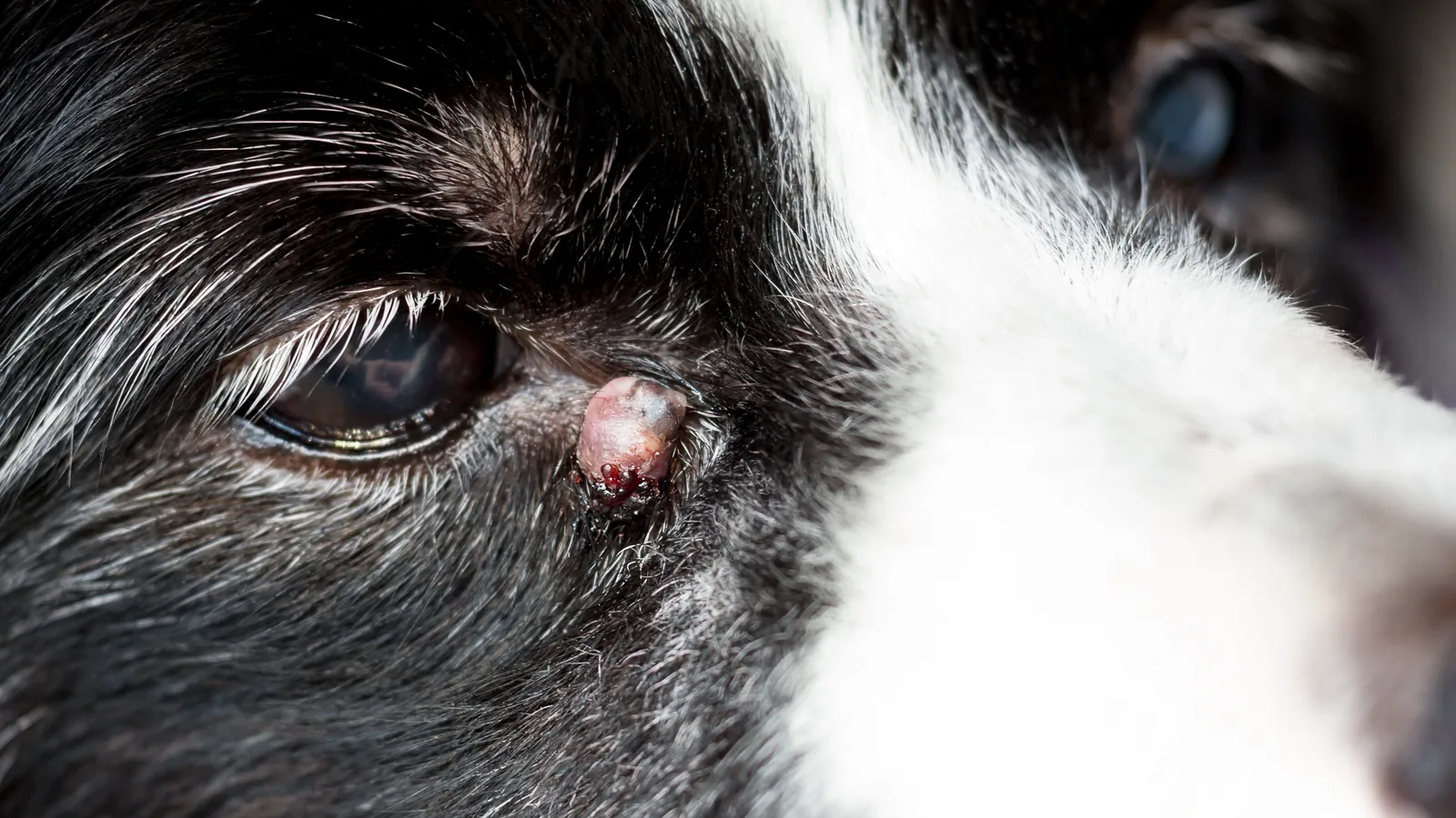
A few things shape how we approach them. They are the most common skin cancer in dogs, making up roughly a fifth of all canine skin tumors, and they are the second most common skin tumor in cats. Because the cells release histamine, the area can swell, redden, and occasionally cause whole-body effects like an upset stomach. Some tumors even seem to grow and shrink on their own. Their appearance is the real challenge, since they can show up as small raised bumps, flat patches, angry ulcerated masses, or growths that look exactly like a harmless fatty lump or a wart. That is exactly why we never judge a lump by how it looks or feels alone.
Which Dogs Are Most at Risk?
Some breeds develop mast cell tumors more often than others. Flat-faced breeds like Boxers, English and French Bulldogs, Pugs, and Boston Terriers sit at higher risk, as do Golden Retrievers, Labradors, Cocker Spaniels, Schnauzers, Beagles, Shar-Peis, and Weimaraners.
That said, any dog can develop one, and most appear in middle-aged to older dogs, though younger dogs are not immune. Regular wellness exams for your dog and simple at-home checks for new or changing lumps are how most of these are caught early, when outcomes are best.
How Do I Recognize a Mast Cell Tumor?
What to Watch for at Home
During monthly lump checks, keep an eye out for:
- Small, movable lumps under the skin.
- Raised bumps that look like insect bites.
- Larger, inflamed, or ulcerated masses.
- Lumps that change size over days or weeks.
- Lumps that turn red, swollen, or painful off and on.
- Hair loss over a lump.
- Itching or licking at one spot.
Less often, a tumor releasing histamine can cause vomiting (sometimes with blood), a poor appetite, diarrhea, or low energy. Monthly checks at home catch most lumps while they are small, and any new or changing lump deserves a veterinary look, because you simply cannot tell from the outside whether a lump is harmless or not.
Telling It Apart From Other Lumps
Several everyday lumps get confused with mast cell tumors. Soft, movable fatty lumps (lipomas) are usually harmless, but that resemblance is exactly why every lump deserves a quick needle test. "It feels like a fatty lump" is not a diagnosis. Fluid-filled cysts can rupture or get infected, and histiocytomas, benign growths common in young dogs, often go away on their own but should be confirmed first.
Getting the diagnosis right matters because the treatments are completely different: a fatty lump can simply be watched, while a mast cell tumor needs surgery with careful margins. Since you cannot reliably tell them apart by look or feel, a fine-needle sample is the standard first step for any new lump.
How Are Mast Cell Tumors Diagnosed?
The First Step: A Needle Sample
A needle sample (fine-needle aspiration) is usually the first test. A small needle draws cells from the lump onto a slide for a look under the microscope. It takes only seconds, rarely needs sedation, is inexpensive, and often gives a quick answer, since mast cells have distinctive granules that are usually easy to spot.
It does have limits. It cannot tell us the tumor's grade, which needs an actual tissue sample, and occasionally the needle misses the tumor cells and gives a false "all clear." Our in-house laboratory runs these samples on site, so most visits come with same-day answers.
Grading: How the Biopsy Guides the Plan
Once a mast cell tumor is confirmed, a surgical biopsy (examining the removed tissue under the microscope) determines its grade, which is the key to predicting how it will behave. You may see grading described on your dog's pathology report with systems like Patnaik (1 to 3) or Kiupel (low versus high), but the practical takeaway is whether the tumor is low, intermediate, or high grade.
| Grade | What it usually means |
| Low-grade | Often cured by surgery alone when removed with clean margins, with cure rates above 90% |
| Intermediate | Behavior varies; a few extra lab tests help predict how it will act |
| High-grade | More likely to spread; usually needs treatment beyond surgery |
Removing the tumor with a wide margin of healthy tissue all around it, plus a layer beneath it, is the strongest predictor of a good long-term outcome. The pathology report also tells us whether those margins were complete, which guides whether any further treatment is needed.
Why Does Checking for Spread Matter?
Staging, the process of checking whether the cancer has spread beyond the original lump, helps us choose the right treatment and set realistic expectations. It looks at the nearby lymph nodes and internal organs, and the results influence whether surgery alone is enough or whether additional treatment makes sense.
| Stage | What it means |
| Stage 0 | Tumor not fully removed, no other spread found |
| Stage I | One tumor, no lymph node involvement |
| Stage II | One tumor with nearby lymph node involvement |
| Stage III | Multiple or large tumors, with or without node involvement |
| Stage IV | Spread to distant sites |
Earlier stages generally carry a better outlook, but later stages still have real treatment options. The conversation just shifts more toward goals and comfort.
What the Workup Involves
A staging workup usually starts with feeling all the lymph nodes and sampling the ones nearest the tumor, even when they feel normal. An abdominal ultrasound checks the spleen, liver, and belly lymph nodes, and chest X-rays look for any spread to the lungs. Our digital X-ray and ultrasound handle this in-house, and bloodwork rounds out the picture by checking your dog's overall health. Not every test is needed for every tumor, so we tailor the plan to your dog's grade, tumor location, and situation.
How Are Mast Cell Tumors Treated?
There are a number of approaches to treating mast cell tumors, depending on their grade and location. Some of these treatments can be done at Noah’s Ark Veterinary Hospital, and others require referral to a veterinary oncologist. We’ll go over the options with you and help you make the best decisions for your pet.
Surgery and Margins
Surgery is the main treatment for most mast cell tumors, and the goal is wide margins. These tumors send tiny, invisible fingers of cells into the surrounding tissue, so removing only the visible lump almost guarantees it grows back. Clean margins on the first surgery dramatically lower that risk, and a repeat surgery in the same spot is harder and less reliable, which is why getting it right the first time matters so much. Our canine surgery services include these wide-margin removals.
Where the tumor sits affects the plan. Tumors on the body often have room for a generous margin, while those on the lower legs are tighter, and for aggressive ones there, amputation occasionally comes up. Face and groin tumors take extra planning. Afterward, care focuses on watching the incision, managing pain, and keeping an eye out for new lumps elsewhere.
Radiation and Chemotherapy
When a tumor cannot be removed with wide margins because of where it sits, radiation therapy can target the microscopic cells left behind and lower the chance of regrowth, though it requires a referral to a specialty oncology center for a multi-week course. Chemotherapy comes into play for high-grade tumors, tumors that have spread to lymph nodes or beyond, or multiple tumors at once. For high-grade or advanced cases, combining surgery with chemo or radiation is often the standard approach.
Supportive Medications
Mast cells release histamine and other irritating chemicals when they break open, which can cause inflammation, stomach ulcers, and occasionally clotting problems. To manage this, we often use antihistamines and stomach protectants like famotidine and omeprazole, and sometimes steroids. Giving antihistamines before surgery lowers the risk of a reaction, and dogs with multiple or recurring tumors may stay on these medications longer term.
What Newer Treatments Are Available Beyond Surgery?
Beyond the traditional trio of surgery, radiation, and chemotherapy, several newer treatments have changed what is possible for mast cell tumors. Some dissolve a tumor without an operation, others recruit the immune system or target the specific signals driving the cancer, and clinical trials open the door to the latest options. We can help you decide which, if any, fit your dog.
Stelfonta: An Injection That Dissolves the Tumor
Stelfonta is an injectable drug placed directly into a mast cell tumor that has not spread. It works by activating a protein (protein kinase C) that causes the tumor to break apart and die, often within days, with the site healing over the following weeks. It is a useful option for tumors that are hard to remove surgically or when an operation is not the right fit, and it spares the dog surgery altogether. Because it triggers histamine release as the tumor breaks down, dogs are pre-treated with antihistamines and steroids.
Gilvetmab: Harnessing the Immune System
Gilvetmab is a newer monoclonal antibody, a type of immunotherapy that helps a dog's own immune system recognize and attack cancer cells, much like some of the immunotherapies used in human medicine. It is given as an injection and works very differently from chemotherapy, which makes it a promising option in certain cases, either alone or alongside other treatments. As a newer, conditionally licensed therapy, it is something we discuss case by case based on your dog's tumor and overall health.
Targeted Therapy (Tyrosine Kinase Inhibitors)
Some mast cell tumors are driven by a mutation in a gene called c-Kit, and targeted oral drugs called tyrosine kinase inhibitors can block that growth signal. Options include toceranib (Palladia), masitinib, and imatinib. They are typically used for tumors that have spread, cannot be fully removed, or have come back, and a tumor can be tested for the c-Kit mutation to predict how well it will respond. These are given as pills at home, with regular monitoring for side effects.
Clinical Trials
For dogs with advanced tumors or disease that is not responding to standard care, clinical trials can offer access to cutting-edge treatments before they are widely available, sometimes at reduced cost. Universities and specialty oncology centers run these studies to test new drugs and combinations. We are glad to help you look into whether a trial might be a good fit for your dog's specific situation.
What Is the Prognosis for a Mast Cell Tumor?
What Shapes the Outlook
A dog's outlook depends on several things working together, and many dogs do very well:
- Grade: low-grade tumors removed completely are often cured, with cure rates above 90%, while high-grade tumors are less predictable.
- Whether it has spread: disease still confined to the original lump carries a better outlook than disease that has reached the lymph nodes or beyond.
- Location: a few spots, like the groin and lower limbs, have historically been trickier, though modern treatment has improved this.
- Clean margins: removing the whole tumor on the first surgery substantially improves the outlook.
Many dogs with low-grade, fully removed tumors are cured with surgery alone and go on to live full lives. High-grade or spread tumors carry a more guarded outlook, but treatment can still give a good quality of life for meaningful stretches of time.
Monitoring After Treatment
After treatment, we recheck your dog regularly, usually every three months for the first year, then every six months, with repeat staging tests as needed. Just as important are your eyes and hands at home, since checking for new lumps anywhere on the body catches problems early, the same way the first tumor was found. Any change at the surgery site or a new lump elsewhere is worth a prompt call. Dogs that have had one mast cell tumor are more likely to develop another, so steady monitoring pays off.
Frequently Asked Questions About Mast Cell Tumors
Can a mast cell tumor be benign?
Low-grade mast cell tumors behave benignly in most cases when completely removed, with cure rates above 90% in some studies. They are still cancer, since the cells are malignant by nature, but the practical outcome with good treatment is often a single surgery and ongoing monitoring rather than a long disease course. Grade matters more than the label.
My dog had a mast cell tumor removed last year. Should I worry about new lumps?
Keep up the monthly checks, but try not to live in fear of the next one. Dogs that have had one mast cell tumor are more likely to develop additional tumors, which is not the same as the first one coming back. Monthly checks at home and steady recheck appointments catch new lumps while they are still small.
What about cats with mast cell tumors?
Cats develop both skin and internal (most often splenic and intestinal) mast cell tumors. Skin tumors in cats often behave more gently than in dogs, while internal disease is more concerning and usually involves a specialist. The overall approach is similar, but the specifics differ, so we talk through feline cases one at a time.
Catching It Early, Treating It Thoroughly
Hearing the words "mast cell tumor" is hard, but most dogs with these tumors do well when the workup and treatment are thorough from the start. We are here to walk through diagnosis, checking for spread, surgery, and follow-up with you, one step at a time. If you have noticed a new lump or you want a second look at one we already know about, request an appointment and we will get your pet on the schedule.